Hypophosphatemic Rickets
Hypophosphatemic Rickets
Also known as:Hypophosphatemic Rickets, X-linked Hypophosphatemia (XLH)
Hypophosphatemic rickets is a group of inherited or acquired diseases in which excessive phosphate loss by the kidneys leads to low blood phosphate levels, causing impaired bone mineralization; it presents as rickets in children and osteomalacia in adults.
Start Here
A quick guide to the next step: which department to start with, what to prepare, and what to ask.
For children, it is recommended to first visit a pediatric endocrinology or pediatric genetics/metabolism department; adults can visit an endocrinology, bone metabolism, or nephrology department. Orthopedics can provide evaluation if there are bone deformities or mobility limitations. Some patients may consider multidisciplinary (MDT) clinics.
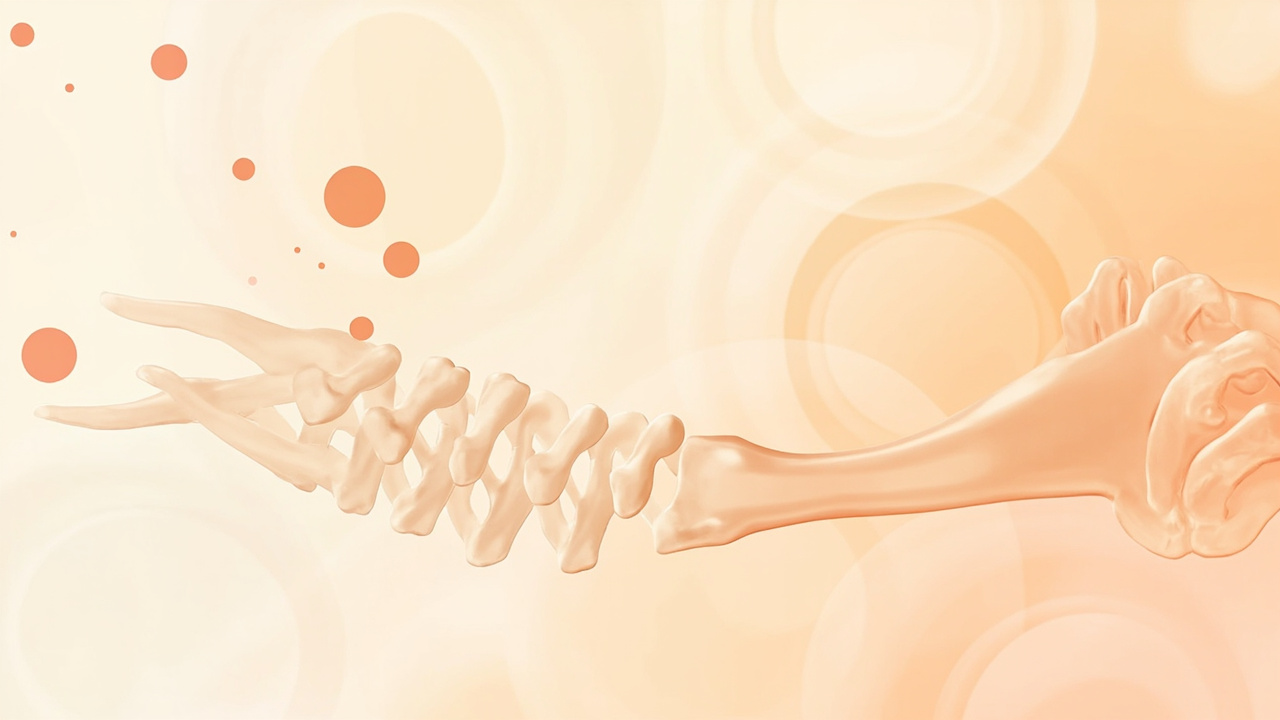
Hypophosphatemic rickets is a group of bone mineralization disorders characterized by low blood phosphate levels. Mutations in genes such as PHEX, FGF23, and DMP1, or acquired causes, lead to abnormal levels of phosphate-regulating factor fibroblast growth factor 23 (FGF23), reduced kidney reabsorption of phosphate, low blood phosphate, and impaired bone mineralization. In children, it manifests as rickets (square head, pigeon chest, O-leg or X-leg deformities, etc.); in adults, it manifests as osteomalacia (fatigue, bone pain, multiple fractures, shortened height).
There is long-term management, including phosphate metabolism monitoring, traditional treatment or targeted therapy evaluation, orthopedic and dental follow-up. Whether specific treatment is suitable must be determined by pediatric/adult endocrinology, nephrology, or bone metabolism specialists.
Yes, inherited hypophosphatemic rickets is mostly X-linked dominant (XLH, caused by PHEX gene mutations), with autosomal dominant/recessive forms also existing. Acquired hypophosphatemic osteomalacia is most commonly tumor-related (TIO).
The presentation of hypophosphatemic rickets is similar to nutritional rickets; primary care physicians often lack awareness of inherited rickets and may misdiagnose it as ordinary vitamin D deficiency rickets, leading to delayed appropriate treatment. Some patients with milder symptoms are only diagnosed in adulthood.
This page helps patients and families organize care leads. It does not replace a clinician’s diagnosis or treatment plan. For testing, medication, referrals, emergency care, and support applications, follow qualified clinicians, medical institutions, support organizations, and official sources.
Diagnosis Path
Organized around the practical patient journey: identify clues, avoid common delays, then prepare for care.
When to Suspect It
- Bone deformities such as square head, pigeon chest, rib beading, and leg bowing (O-leg or X-leg) in childhood
- Slowed growth in height; X-ray of long bones showing widened, blurred metaphyses with frayed cupping
- Persistently low blood phosphate, significantly elevated blood alkaline phosphatase, but normal blood calcium, vitamin D, and parathyroid hormone levels
- Family history of rickets or osteomalacia
- Fatigue, bone pain, multiple pseudofractures, and gradual height loss in adults
- Poor response to standard calcium or vitamin D supplementation for rickets
Common Wrong Turns
- Misdiagnosis as nutritional vitamin D deficiency rickets based solely on clinical presentation, with only calcium or regular vitamin D supplementation
- Neglecting to take a detailed family history and perform genetic testing to identify the underlying cause
- Failure to systematically differentiate causes of hypophosphatemia, missing inherited or tumor-related possibilities
- For acquired hypophosphatemic osteomalacia, failing to actively search for tumor-related clues, delaying treatment of the underlying cause
Departments to Start With
- Pediatric endocrinology or pediatric genetics/metabolism (children)
- Endocrinology or bone metabolism (adults)
- Orthopedics (significant bone deformities or surgical evaluation needed)
- Nephrology (suspected kidney disease causing phosphate metabolism abnormalities)
- Pediatric urology/andrology or reproductive endocrinology (for gonadal function assessment)
Before the Visit
- Detailed medical history: age of onset, growth and development history, family history
- Serum biochemical tests: blood phosphate, calcium, alkaline phosphatase, serum FGF23, 25-hydroxyvitamin D, parathyroid hormone
- Urine phosphate test: calculate tubular phosphate reabsorption rate (TmP/GFR)
- Imaging: X-ray of long bones (assess rickets changes), bone CT/MRI (if needed)
- Genetic testing: for suspected inherited hypophosphatemic rickets, test genes such as PHEX, FGF23, DMP1, ENPP1, SLC34A3
- Tumor screening: for suspected TIO, perform imaging to locate the causative tumor
Tests to Ask About
- Serum phosphate, calcium, alkaline phosphatase
- 25-hydroxyvitamin D and parathyroid hormone
- Urine phosphate and tubular phosphate reabsorption rate
- Serum FGF23 level
- X-ray of long bones
- Genetic testing (PHEX, etc.)
Questions for the Doctor
- Are my child's bone deformities due to hypophosphatemic rickets or ordinary nutritional rickets? What tests are needed for diagnosis?
- Does my child need genetic testing? Should other children in the family be tested?
- What long-term medications will my child need? What are the side effects?
- What should my child eat? Should activity be restricted?
- Do I need to continue treatment and follow-up after reaching adulthood?
- If I want to have another child, do I need prenatal counseling?
Basic Information
Medical Notes
More complete medical explanations are kept here for discussion with clinicians.
Symptoms
Diagnosis
Treatment
Long-term Care
Fertility and Family
When to Seek Urgent Care
Care Resources
No verified hospital or department leads are available for this disease yet. Use the starting departments below to prepare, and confirm registration or referral details with clinicians and official hospital channels.